Histopathology department
The Histopathology Department in a laboratory specializes in the examination of tissue specimens to diagnose diseases, particularly cancer and other conditions affecting organs and tissues. Here’s an overview of what you might find in a typical Histopathology Department:
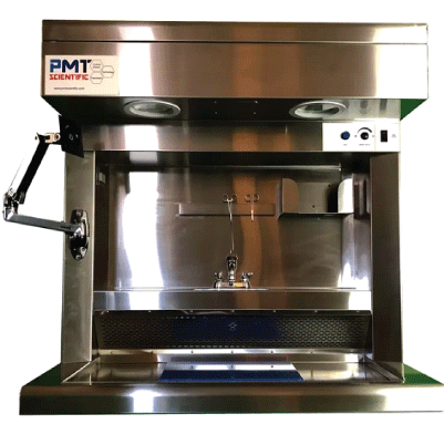
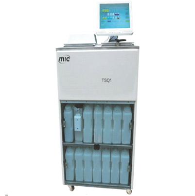
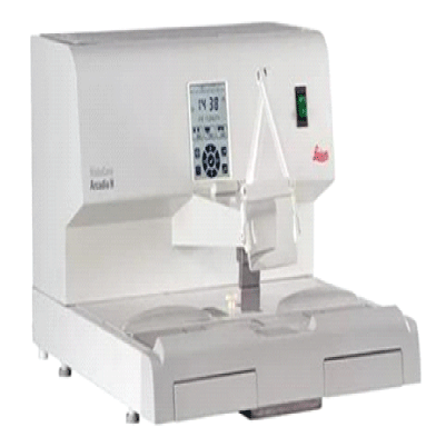
Tissue Processing: Histotechnologists in the department receive tissue specimens from surgical procedures, biopsies, or autopsies. They process these specimens by embedding them in paraffin wax blocks, which preserves the tissue structure for microscopic examination.
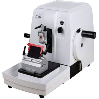
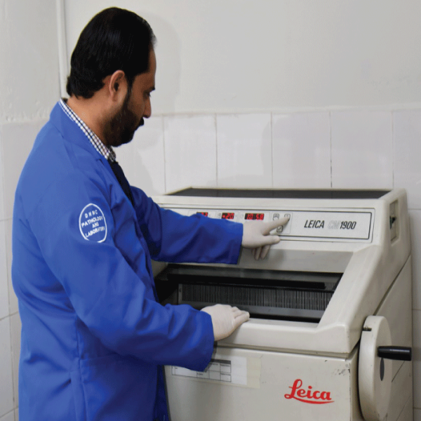
Microtomy: Histotechnologists use microtomes to cut thin sections (slices) of tissue from paraffin blocks. These sections are mounted on glass slides and stained using various techniques to highlight different cellular structures and abnormalities.
Staining Techniques: Different staining techniques are employed to enhance the visibility of specific tissue components and pathological changes. Common stains used in histopathology include hematoxylin and eosin (H&E) stain, which provides contrast between cell nuclei and cytoplasm, and special stains for highlighting particular structures or substances (e.g., immunohistochemistry, silver stains).
Slide Preparation: Once stained, tissue sections are coverslipped to protect them and ensure clarity under the microscope. Histotechnologists may also perform additional processing steps, such as deparaffinization and hydration, to prepare slides for examination.
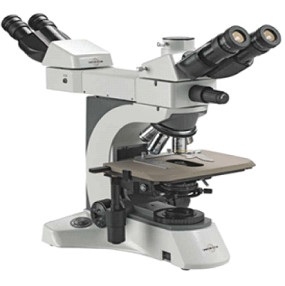
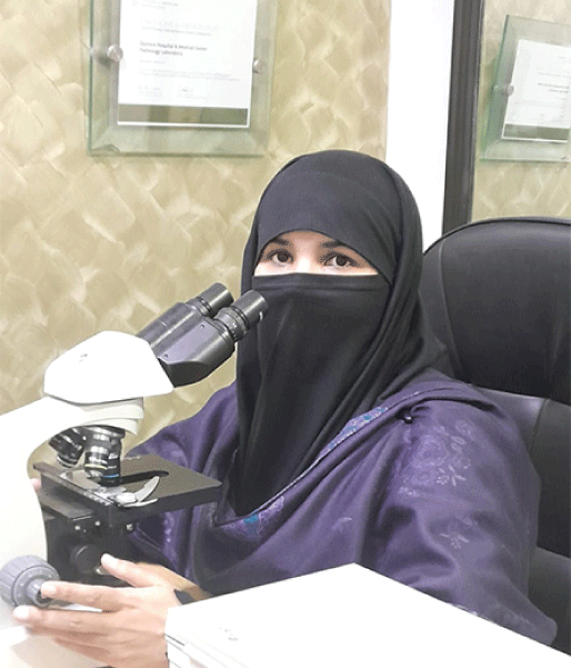
Microscopic Examination: Histopathologists examine stained tissue sections under a microscope to identify abnormal cellular features, tissue architecture, and pathological changes indicative of disease. They correlate these findings with clinical information to make accurate diagnoses and provide valuable prognostic and therapeutic information.
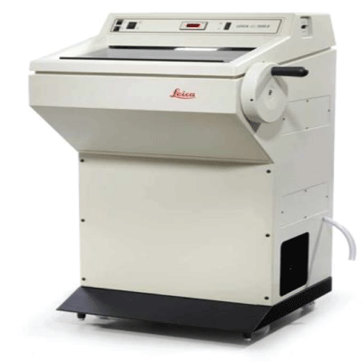
Frozen Section Analysis: In some cases, rapid frozen section analysis is performed during surgical procedures to provide immediate diagnostic information. Histotechnologists quickly freeze tissue samples, cut thin sections, and stain them for rapid evaluation by a pathologist, allowing surgeons to make real-time decisions regarding patient care.
Specialized Techniques: Histopathology departments may utilize specialized techniques to analyze tissue specimens in greater detail. This includes immunohistochemistry, which involves labeling specific proteins or antigens in tissue sections to aid in diagnosis and classification of tumors, as well as molecular testing to detect genetic mutations, gene rearrangements, and other molecular alterations associated with cancer.
Consultation and Collaboration: Histopathologists often collaborate with other healthcare providers, including surgeons, oncologists, and radiologists, to ensure accurate diagnosis and appropriate patient management. They may also consult with experts in subspecialty areas of pathology for challenging cases or rare diseases.
Quality Assurance: Quality assurance measures are integral to histopathology practice to maintain the accuracy and reliability of diagnostic interpretations. This includes adherence to standardized protocols and guidelines, ongoing training and competency assessment for laboratory staff, participation in proficiency testing programs, and regular review of diagnostic discrepancies.
Overall, the Histopathology Department plays a crucial role in the diagnosis, staging, and management of diseases through the examination of tissue specimens, contributing to improved patient outcomes and treatment decisions.

